
Hip Treatment, Dr Thomas Youm, Orthopaedic Hip Surgeon in New York, NY and New Jersey
Hip Anatomy
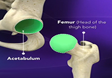
The hip joint is the largest weight-bearing joint in the human body. It is also referred to as a ball and socket joint and is surrounded by muscles, ligaments and tendons. The thighbone or femur and the pelvis join to form the hip joint.
Osteoarthritis of the Hip
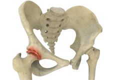
Osteoarthritis, also called degenerative joint disease, is the most common form of arthritis. It occurs most often in older people. This disease affects the tissue covering the ends of bones in a joint (cartilage).
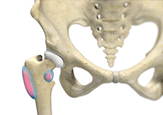
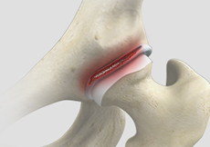
Hip Labral Tears
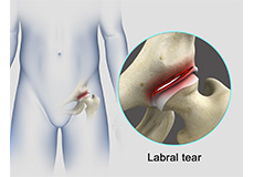
A hip labral tear is an injury to the labrum, the cartilage that surrounds the outside rim of your hip joint socket.
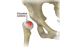
Chondral Lesions or Injuries
The hip joint is one of the largest weight-bearing joints in the body, formed by the thighbone or femur and the acetabulum of the pelvis. It is a ball and socket joint with the head of the femur as the ball and the pelvic acetabulum forming the socket. The joint surface is covered by a smooth articular cartilage which acts as a cushion and enables smooth movements of the joint.
Loose Bodies
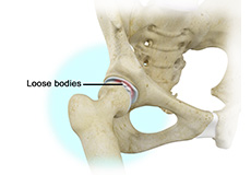
Loose bodies are small loose fragments of cartilage or a bone that float around the joint. The loose bodies can cause pain, swelling, locking and catching of the joint.
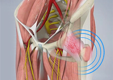
Hip Synovitis
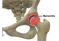
Hip synovitis, also called transient hip synovitis or toxic synovitis, is a condition in which there is inflammation of the synovial tissues surrounding the hip joint causing hip pain. It is the most common reason for sudden hip pain occurring in young children between the age of 2 and 9.
Hip Instability
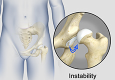
The hip plays an important role in supporting the upper body weight while standing, walking and running, and hip stability is crucial for these functions. The femur (thighbone) and acetabulum (hip bone) join to form the hip joint, while the labrum (tissue rim that seals the hip joint) and the ligaments lining the hip capsule maintain the stability of the hip.
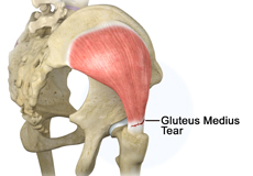
Gluteus Medius Tear
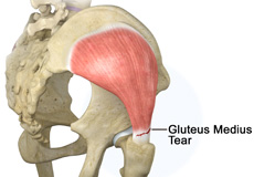
A gluteus medius tear is a condition characterized by severe strain on the gluteus medius muscle that results in partial or complete rupture of the muscle.
The gluteus medius is one of the major muscles of the hip and is essential for movement of the lower body and keeping the pelvis level during ambulation.
Trochanteric Bursitis
Hip bursitis is a painful condition caused by inflammation of a bursa in the hip. Bursae are fluid- filled sacs present in joints between bone and soft tissue to reduce friction and provide cushioning during movement.
Snapping Hip Syndrome
The hip is an important joint that helps us walk, run and jump. The ball-and-socket joint in the hip is formed between the round end of the femur (thighbone) and the cup-shaped socket of the acetabulum (part of the hip bone).
Hip Arthroscopy
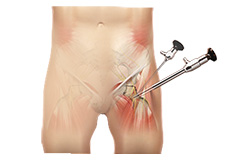
This information sheet is aimed to provide a general guide to the common indications for hip arthroscopy, what to expect if undergoing the surgery, the significant risks, and a general guide to the expected recovery pathway. This document is not by any means fully comprehensive, and any particular questions should be addressed with your surgeon. Many different hip procedures can be performed arthroscopically, and from country to country, and surgeon to surgeon, there will be significant variations.
Arthroscopic Assisted Core Decompression of the Hip
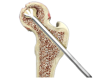
While arthroscopy has been available to the orthopaedic community since its development in 1931, hip arthroscopy has only recently become popularized. A growing number of indications for hip arthroscopy has caused the number of procedures to increase eighteen-fold between 1999 and 2009.4 Current indications for hip arthroscopy include symptomatic loose bodies, labral lesions, femoral acetabular impingement (FAI), acute articular injury, traumatic rupture of the ligamentum teres, instability, peritrochanteric disorders (snapping hip, bursitis, gluteus medius tears), and joint sepsis.2 Yet another indication for hip arthroscopy is arthroscopic-assisted core decompression for avascular necrosis (AVN) of the femoral head.
Arthroscopic Gluteus Medius Tendon Repair
Coming soon
Hamstring Repair & Reconstruction
The three hamstring muscles, namely semitendinosus, semimembranosus and biceps femoris, run down the back of the thigh and help you bend (flex) your knee and extend your leg. Hamstring injuries are common in athletes who participate in sports which involve running such as track, soccer, and basketball.
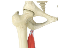
Gluteus Maximus Transfer
Gluteus maximus transfer, also referred to as gluteus maximus flap transfer (GMT), is a surgical technique to treat severe pain, limp, and instability associated with loss of the abductor portions of the gluteus medius and gluteus minimus muscles of the hip secondary to total hip arthroplasty or primary abductor disruption.
Femoral Derotational Osteotomy
Coming soon
Hip Labral Repair
Labrum is a ring of strong fibrocartilaginous tissue lining around the socket of the hip joint. Labrum serves many functions where it acts as shock absorber, lubricates the joint, and distributes the pressure equally. It holds the head of the femur in place and prevents the lateral and vertical movement of the femur head within the joint. It also deepens the acetabular cavity and offers stability against femoral head translation.
Hip Labral Reconstruction
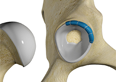
Hip labral reconstruction is a surgery that involves the use of a graft to replace the damaged portion of a hip labrum. The hip labrum is a ring of fibrous cartilaginous tissue that surrounds the socket of the hip joint. Injuries to the hip labrum are usually seen in athletes involved in high-impact sports such as ice hockey, soccer, and football. They can also occur due to traumatic injury or degenerative conditions.